How Do Bacteria Repair Uv Damage

(Phys.org) —From bacteria to plants to humans, all organisms take mechanisms that they use to repair Deoxyribonucleic acid damaged by ultraviolet (UV) light. This key maintenance function is critical to our health because damaged DNA tin can atomic number 82 to diseases such as cancer. So, of class, we know all about how it works. Or so scientists thought. New research has provided evidence that the current model for how UV repair functions must to be reworked.
At consequence is the number of molecules of the important proteins in the complex, UvrA and UvrB. At issue is the number of molecules of the of import proteins in the complex, UvrA and UvrB. This new study, conducted past researchers from Harvard Academy utilizing the U.S. Department of Free energy Office of Science's Advanced Photon Source promises to provide new insights into the fundamental mechanisms of DNA repair and into diseases that are acquired past mutations in these genes such as xeroderma pigmentosum (an autosomal recessive genetic disorder of DNA repair in which the ability to repair damage caused by ultraviolet (UV) lite is deficient), Cockayne syndrome (a disorder characterized by short stature and an appearance of premature aging), and trichothiodystrophy (an inherited condition in which hair is brittle, sparse, and easily cleaved).
Previous work had shown that the bacterial DNA repair mechanism involved three proteins, UvrA, UvrB, and UvrC. The model was that ii copies of UvrA traveled around with one copy of UvrB (A2B1) and scanned the DNA for places where damage had occurred. When a spot was identified that needed repair, the A2B1 stopped and waited at the spot that needed repair. At this point UvrC would come in and displace UvrA and class a circuitous with UvrB (B1C1) that conducted the repair. This model explained how the complex could scan both Dna strands only and then orient to repair only i strand. The asymmetric A2B1 fit perfectly.
The but problem was, data from various structural methods started to pile up that were not consequent with this model and suggested that the initial complex might actually be A2B2. In addition, the Harvard enquiry team solved the crystal construction of the UvrA/UvrB complex and it was consistent with the A2B2 model. They proposed that A2B2 identified the impairment and then the UvrA and one of the UvrB molecules were released when UvrC arrived to repair the problem. Withal, outstanding questions remained about whether there might be other configurations in the crystal structure consistent with the longstanding model.
The group decided the all-time style to answer these questions was to evaluate the complex using small-angle ten-ray scattering (SAXS). Their thinking was that low-resolution structural data collected on the poly peptide circuitous in solution would be ideal for comparing modeling of various protein configurations. In improver, they hoped to find evidence for structural changes associated with how these proteins bind to ATP and use its free energy for their activities.
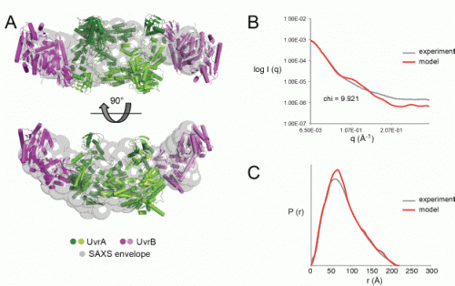
SAXS analysis of a solution of UvrA and UvrB in circuitous carried out at the Biophysics Collaborative Access Team (Bio-Cat) beamline 18-ID-D at the Argonne Avant-garde Photon Source showed an elongated structure (run into the figure).
Modeling of the SAXS data against v possible configurations, four A2B2 options and ane A2B1 option, showed that the information was consistent with one of the A2B2 configurations observed in the crystal structure but ruled out the others. Further assay of the SAXS data by five other methods also supported this decision.
Unfortunately, when the group added ATP or ADP to the complex, they were not able to meet any meaning changes. This may crave further experiments with different versions of the protein that allow them to keep the proteins bound to either ATP or ADP. For now, the Harvard researchers will be working on their new model to explain how leaner perform this universal role.
More than information: Danaya Pakotiprapha, David Jeruzalmi. "Modest-angle X-ray scattering reveals architecture and A2B2 stoichiometry of the UvrA–UvrB Deoxyribonucleic acid impairment sensor," Proteins 81,132 (2013). DOI: 10.1002/prot.24170
Citation: How practise bacteria repair damage from the sunday? (2014, January 24) retrieved 12 April 2022 from https://phys.org/news/2014-01-bacteria-sunday.html
This certificate is subject to copyright. Apart from any fair dealing for the purpose of individual study or research, no part may be reproduced without the written permission. The content is provided for data purposes only.
How Do Bacteria Repair Uv Damage,
Source: https://phys.org/news/2014-01-bacteria-sun.html
Posted by: blountspaccur.blogspot.com


0 Response to "How Do Bacteria Repair Uv Damage"
Post a Comment